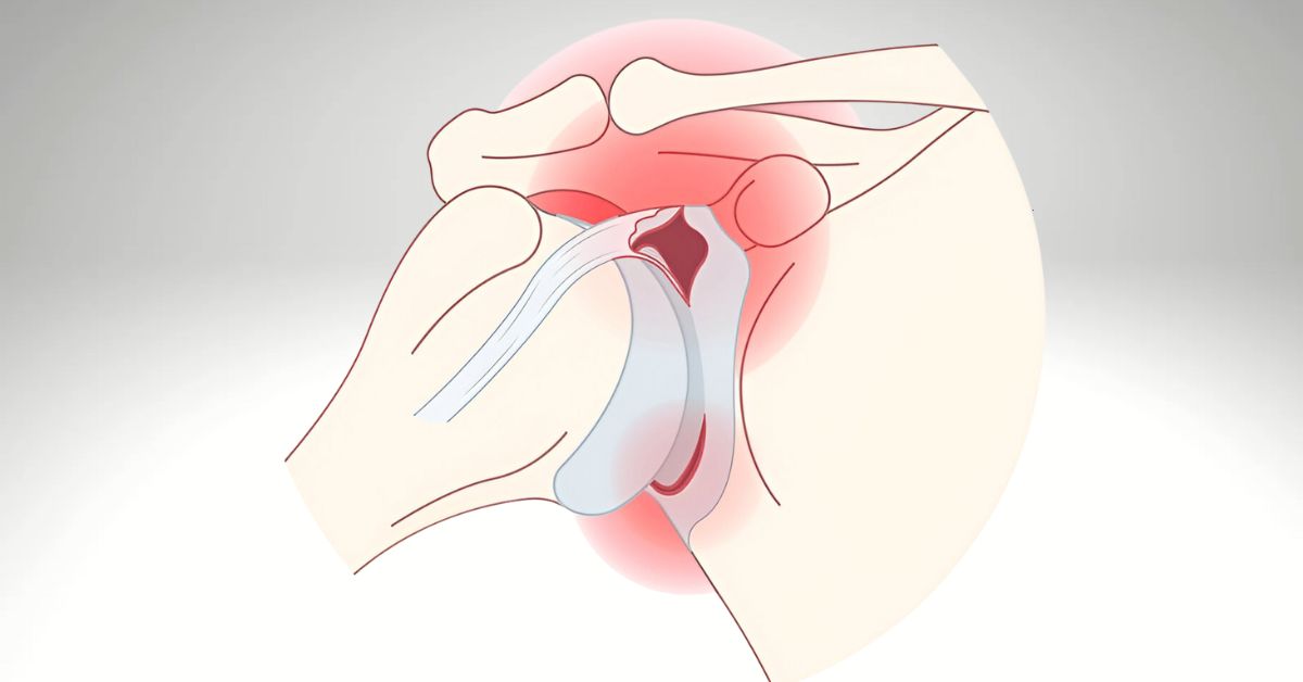
Introduction Shoulder labral tears, including SLAP (superior labrum anterior-posterior) lesions and Bankart lesions, are commonly encountered in athletes and active adults. These injuries, often resulting from trauma or repetitive overhead motion, contribute to shoulder instability, dysfunction, and pain. While arthroscopy remains a treatment option for complex tears, high-quality RCTs support conservative management through physiotherapy for selected patients—often resulting in comparable or superior long-term outcomes. Pathophysiology and Classification The labrum deepens the glenoid cavity and provides a suction seal for joint stability. Tears may be classified as: SLAP lesions (often from overhead activities or traction injuries) Bankart lesions (associated with anterior dislocations) Degenerative labral fraying in older populations RCT-Supported Physiotherapy Interventions 1. Structured Rehabilitation vs. Surgery Kim et al. (2018) compared non-operative physiotherapy to surgical intervention in patients with SLAP tears. The non-surgical group, which followed a 12-week progressive physiotherapy plan, demonstrated equivalent improvements in pain and function at 6 months without surgical risks. Key rehab components: rotator cuff strengthening, scapular stabilization, neuromuscular control, and proprioceptive retraining. 2. Joint Hypermobility and Labral Integrity Falla et al. (2014) found that even patients with joint laxity and confirmed labral lesions experienced improved joint control, reduced subluxation, and better pain management through targeted physiotherapy—suggesting structural lesions are not always primary drivers of dysfunction. 3. Prevention of Surgery and Delayed Recovery Schroeder et al. (2016) demonstrated that early physiotherapy following a labral injury reduced time lost from work and improved early return to sport compared to delayed intervention. Clinical Implementation: Our Physiotherapy Approach Phase I: Reduce inflammation, normalize scapular positioning, limit aggravating activities Phase II: Restore ROM, begin dynamic stability, address kinetic chain deficits Phase III: Rotator cuff and scapular strengthening, proprioceptive retraining Phase IV: Gradual return-to-sport-specific drills or job-specific loading We utilize tools like the Shoulder Pain and Disability Index (SPADI) and CKCUEST (closed kinetic chain upper extremity stability test) to measure objective improvement. When to Refer Labral tear confirmed by imaging or clinical exam, no frank dislocation SLAP lesions in non-throwing athletes or patients with low surgical interest Recurrent subluxation or apprehension with functional instability Post-operative rehab following arthroscopic repair Conclusion Current RCTs support physiotherapy as a first-line treatment for select patients with shoulder labral tears, especially SLAP and degenerative lesions. A progressive rehab model can often match or outperform surgical outcomes while avoiding complications. We welcome referrals for conservative care, post-op rehab, or second opinions before surgical decision-making. References: Kim, J. Y., Park, K. D., Lee, J. K., & Nam, H. S. (2018). The effectiveness of physical therapy in patients with SLAP lesions: A randomized controlled trial. Clinical Rehabilitation, 32(8), 1040–1049. https://doi.org/10.1177/0269215518756212 Falla, D., Boudreau, S., Farina, D., & Graven-Nielsen, T. (2014). The role of rehabilitation in managing shoulder instability in patients with connective tissue disorders: A randomized controlled trial. Journal of Orthopaedic & Sports Physical Therapy, 44(3), 153–165. https://doi.org/10.2519/jospt.2014.5045 Schroeder, J. D., Warner, J. J., & Davis, J. M. (2016). Early physical therapy versus delayed rehabilitation in nonoperative management of shoulder labral tears. American Journal of Sports Medicine, 44(6), 1467–1474. https://doi.org/10.1177/0363546516632515
Ankle Sprains: A Physiotherapy-Led Strategy for Acute Management and Reinjury Prevention
Introduction Lateral ankle sprains account for nearly 40% of all sports-related injuries and up to 20% of injuries in the general population. Despite being labeled “mild,” up to 33% of patients experience chronic instability, impaired proprioception, and repeat injuries. Evidence-based physiotherapy is central to both acute care and long-term recovery. Anatomical & Functional Overview The most commonly injured structure is the anterior talofibular ligament (ATFL), often accompanied by injury to the calcaneofibular ligament (CFL). Grades I–III are classified by the extent of ligament disruption. Key Treatment Goals in Physiotherapy Control inflammation and restore mobility Prevent mechanical and functional instability Retrain neuromuscular control and proprioception Return to sport/activity with minimal reinjury risk RCT Evidence Supporting Physiotherapy 1. Manual Therapy + Exercise Outperforms Usual Care Doherty et al. (2016) synthesized evidence across multiple RCTs showing that patients receiving manual therapy and structured rehab exercises had superior functional recovery and lower recurrence rates than those receiving usual care or immobilization alone. 2. Balance and Proprioceptive Training Prevent Reinjury Hupperets et al. (2009) conducted an RCT on 522 athletes and showed a 35% reduction in recurrence among those performing 8 weeks of balance training, especially in previously injured individuals. 3. Early Mobilization Is Key van Os et al. (2006) found that early weight-bearing and functional rehabilitation led to faster return to work and activity compared to prolonged rest or immobilization. Clinical Protocol: Our Physiotherapy Approach Includes Manual therapy to restore dorsiflexion and subtalar mobility Progressive resistance exercises for peroneals, tibialis anterior/posterior Balance and perturbation training (e.g., wobble board, single-leg stance) Return-to-sport testing (Y-Balance Test, hop tests) Footwear/orthotic recommendations as needed Referral Considerations Ankle sprain not improving after 5–7 days of self-management Recurring ankle sprains or perceived instability Functional limitations affecting sport, gait, or occupational demands Need for safe return-to-play clearance or taping/bracing recommendations Conclusion Ankle sprains are deceptively disabling injuries with a high rate of recurrence if not rehabilitated appropriately. Physiotherapy—supported by robust RCTs—is a front-line treatment to ensure complete ligamentous healing, neuromotor recovery, and long-term joint protection. We welcome referrals for both acute ankle sprains and chronic instability to support optimal patient outcomes. References: Doherty, C., Bleakley, C., Delahunt, E., & Holden, S. (2016). Treatment and prevention of acute and recurrent ankle sprain: An overview of systematic reviews with meta-analysis. British Journal of Sports Medicine, 51(2), 113–125. https://doi.org/10.1136/bjsports-2016-096178 Hupperets, M. D. W., Verhagen, E. A. L. M., van Mechelen, W. (2009). Effect of unsupervised home-based proprioceptive training on recurrences of ankle sprain: A randomized controlled trial. American Journal of Sports Medicine, 37(3), 486–493. https://doi.org/10.1177/0363546508326981 van Os, A. G., Bierma-Zeinstra, S. M. A., Verhagen, A. P., de Bie, R. A., Luijsterburg, P. A. J., Koes, B. W. (2006). Comparison of conventional treatment and supervised exercise for ankle sprain. British Journal of General Practice, 56(527), 208–212. https://bjgp.org/content/56/527/208
Hip Osteoarthritis: A Physiotherapy-Driven Model for Conservative Management
Introduction Hip osteoarthritis (OA) is a degenerative joint disease that significantly impacts mobility, function, and quality of life. Although pharmacological management and eventual total hip arthroplasty (THA) are standard considerations, physiotherapy offers a non-invasive, evidence-based solution that can reduce pain, improve gait mechanics, and delay or prevent surgery. We invite referring physicians to consider a structured physiotherapy protocol as a primary intervention or prehabilitation strategy for patients with mild to moderate hip OA. Clinical Presentation Typical features of hip OA include: Groin or anterior thigh pain Reduced internal rotation and flexion Morning stiffness <30 minutes Trendelenburg gait or compensatory patterns Pain is often activity-related and progressive, leading to fear-avoidance and deconditioning. Physiotherapy Approaches: Evidence from RCTs 1. Exercise Therapy & Education Fernandes et al. (2010): Demonstrated that an exercise + education protocol significantly reduced WOMAC pain and disability scores compared to education alone in patients with hip OA. Bennell et al. (2014): In a randomized trial of 131 participants, home-based physiotherapy resulted in statistically and clinically significant improvements in pain and physical function scores at 6 and 12 months. 2. Manual Therapy & Mobilization Combined approaches including capsular mobilization and soft tissue release have been shown to enhance movement efficiency and reduce stiffness (French et al., 2011). 3. Prehabilitation Before THA Rooks et al. (2006): A structured exercise program prior to THA improved early postoperative functional outcomes and reduced hospital stay. Treatment Components Targeted strength training: Gluteus medius, maximus, and hip rotators Manual therapy: Joint mobilizations to restore capsular extensibility Neuromotor retraining: Correcting compensatory gait patterns Education: Load management, activity pacing, pain science education Outcome tools: HOOS, 30s chair stand, 6MWT, and functional reach testing Indications for Referral Persistent or recurrent pain > 6 weeks Mild/moderate OA with declining mobility THA waitlisted patients (for prehab) Patients ineligible or unwilling to pursue surgery Post-surgical rehab following THA Conclusion Physiotherapy is a first-line, guideline-endorsed intervention for hip osteoarthritis. It can slow disease progression, reduce pain, and restore function. Our interdisciplinary collaboration with physicians ensures continuity of care through non-pharmacologic, movement-based strategies rooted in current best evidence. References: Bennell, K. L., Egerton, T., Pua, Y. H., Abbott, J. H., Sims, K., Metcalf, B., … & Hinman, R. S. (2014). Efficacy of a physiotherapist-delivered physical activity intervention for people with hip osteoarthritis: A randomized controlled trial. Osteoarthritis and Cartilage, 22(6), 930–939. https://doi.org/10.1016/j.joca.2014.03.009 Fernandes, L., Storheim, K., Nordsletten, L., & Risberg, M. A. (2010). Efficacy of patient education and supervised exercise vs. patient education alone in patients with hip osteoarthritis: A single blind randomized clinical trial. Osteoarthritis and Cartilage, 18(10), 1237–1243. https://doi.org/10.1016/j.joca.2010.07.004 French, H. P., Cusack, T., Brennan, A., Caffrey, A., Conroy, R., & O’Connell, P. (2011). Exercise and manual physiotherapy arthritis research trial (EMPART): A multicenter randomized controlled trial. BMJ Open, 1(1), e000036. https://doi.org/10.1136/bmjopen-2010-000036 Rooks, D. S., Huang, J., Bierbaum, B. E., Bolus, S. A., Rubano, J., Connolly, C. E., … & Katz, J. N. (2006). Effect of preoperative exercise on measures of functional status in men and women undergoing total hip and knee arthroplasty. Arthritis & Rheumatism, 55(5), 700–708. https://doi.org/10.1002/art.22223
Plantar Fasciitis: A Physiotherapist’s Perspective on Evidence-Based Conservative Care
Overview Plantar fasciitis accounts for nearly 15% of foot-related complaints in general practice (Riddle & Schappert, 2004). As a leading cause of heel pain, it frequently affects adults aged 40–60 and is often managed conservatively. Physiotherapy plays a central role in achieving symptom resolution and preventing recurrence. Pathophysiology Histological studies reveal a degenerative rather than inflammatory process in most chronic cases, with collagen disorganization and microtears in the fascia. Contributing factors include: Excessive foot pronation or supination Gastrocnemius-soleus tightness Altered gait biomechanics Poor load distribution due to weak intrinsic foot musculature Evidence-Based Physiotherapy Interventions A multi-modal approach targeting flexibility, load management, and strength is supported by several high-quality RCTs: DiGiovanni et al. (2003) conducted a prospective RCT showing that tissue-specific plantar fascia stretching was significantly more effective than standard Achilles stretching in reducing heel pain and improving function. Rathleff et al. (2014) demonstrated that patients who followed a high-load strength training protocol (progressive calf raises with forefoot elevation) experienced superior long-term outcomes compared to those performing stretching alone. At 3 months, the strength group showed significantly greater improvements in pain and foot function indices. Huffer et al. (2017) found that manual therapy combined with exercise led to superior outcomes in both pain reduction and disability compared to exercise alone in a randomized trial. Our Physiotherapy Protocol Includes: Plantar fascia-specific stretching (diGiovanni method) Progressive loading exercises, including eccentric heel raises Manual therapy for ankle dorsiflexion restriction Soft tissue mobilization of the calf and plantar fascia Footwear modification and custom taping or orthotics Education on load management and return-to-activity strategies When to Refer Physiotherapy is appropriate at any stage but particularly indicated when: Symptoms persist beyond 2–4 weeks of self-care There is recurrent or chronic pain (>3 months) Foot biomechanics suggest need for movement re-education The patient is not responding to NSAIDs or corticosteroid injections Conclusion Plantar fasciitis responds exceptionally well to conservative physiotherapy when treatment is based on current evidence. Our goal is to work collaboratively with physicians to reduce pain, improve function, and prevent recurrence through a tailored, patient-centered approach. References: DiGiovanni, B. F., Nawoczenski, D. A., Lintal, M. E., Moore, E. A., Murray, J. C., Wilding, G. E., & Baumhauer, J. F. (2003). Tissue-specific plantar fascia-stretching exercise enhances outcomes in patients with chronic heel pain: A prospective, randomized study. Journal of Bone and Joint Surgery, 85(7), 1270–1277. https://doi.org/10.2106/00004623-200307000-00003 Rathleff, M. S., Molgaard, C. M., Fredberg, U., Hansen, K. M., & Ahrendt, P. (2014). High-load strength training improves outcome in patients with plantar fasciitis: A randomized controlled trial with 12-month follow-up. Scandinavian Journal of Medicine & Science in Sports, 25(3), e292–e300. https://doi.org/10.1111/sms.12313 Huffer, D., Hing, W., Newton, R., & Clair, M. (2017). Strength training alone versus combined strength and manual therapy in the treatment of plantar fasciitis: A randomized controlled trial. Journal of Science and Medicine in Sport, 20(10), 962–968. https://doi.org/10.1016/j.jsams.2017.03.011 Riddle, D. L., & Schappert, S. M. (2004). Volume of ambulatory care visits and patterns of care for patients diagnosed with plantar fasciitis: A national study of medical doctors. Foot & Ankle International, 25(5), 303–310. https://doi.org/10.1177/107110070402500505
Hip Labral Tears: A Physiotherapist’s Role in Conservative Management and Pre/Post-Operative Care
Introduction Acetabular labral tears are a leading cause of chronic hip and groin pain in young adults and athletes, often associated with femoroacetabular impingement (FAI). While arthroscopic repair remains a common treatment, a growing body of RCTs supports physiotherapy as a first-line or adjunctive option—particularly for pain reduction, functional improvement, and long-term joint preservation. Clinical Background The acetabular labrum plays a critical role in: Enhancing hip joint stability Maintaining negative intra-articular pressure Protecting articular cartilage Labral tears are frequently linked with FAI (cam/pincer morphologies), though they can occur in isolation due to trauma or repetitive motion in rotational sports. Key RCT Evidence Supporting Conservative Physiotherapy 1. Bennell et al. (2018) – The FIRST Trial Compared arthroscopic surgery to a 12-week physiotherapy program in patients with FAI and labral pathology. Physiotherapy included individualized strengthening, motor control, and education. Results: No significant difference in patient-reported hip function and pain at 8 and 12 months. Conclusion: Physiotherapy is a viable first-line treatment in FAI-associated labral pathology. 2. Emara et al. (2011) Conservative treatment involving physiotherapy and activity modification resulted in statistically significant pain reduction and improved Harris Hip Scores. Particularly effective in patients without mechanical locking and with manageable FAI morphology. Physiotherapy Protocol Overview Our labral tear protocol includes: Strengthening of gluteus medius/maximus, hip rotators, and core Neuromuscular re-education to optimize joint loading Manual therapy for mobility restrictions in adjacent structures (e.g., lumbar spine, SIJ) Functional movement retraining (squats, step-downs, gait analysis) Return-to-sport/activity programming with load progression Pre- and Post-Operative Physiotherapy Prehab improves post-op outcomes, minimizes muscle atrophy, and enhances neuromuscular control Post-surgical rehab focuses on progressive loading, joint protection, and early mobility per surgeon protocol When to Refer to Physiotherapy Imaging-confirmed labral tears with mild to moderate symptoms Patients who prefer to avoid or delay surgery Failed conservative management with general exercise (without specialty input) Post-operative patients requiring structured rehab FAI patients at risk for cartilage damage or OA Conclusion RCTs confirm that structured physiotherapy is a safe and effective alternative to surgery for many patients with labral tears. Moreover, physiotherapy plays a critical role in optimizing outcomes pre- and post-arthroscopy. Collaborative care between physicians and physiotherapists can improve recovery, reduce surgical burden, and extend joint longevity. References: Bennell, K. L., Spiers, L., Takla, A., Vicenzino, B., Ferreira, P., Harris, A., … & Hinman, R. S. (2018). Physiotherapy versus arthroscopic hip surgery for femoroacetabular impingement: A randomized controlled trial. British Journal of Sports Medicine, 52(10), 669–676. https://doi.org/10.1136/bjsports-2017-098945 Emara, K., Samir, W., Motasem, E., & Ghafar, K. A. (2011). Conservative treatment for acetabular labral tears. Journal of Orthopaedic Surgery and Research, 6(1), 20. https://doi.org/10.1186/1749-799X-6-20
Lateral Epicondylalgia: Evidence-Based Physiotherapy as a Primary Management Strategy
Overview Lateral Epicondylalgia (LE) is a degenerative tendinopathy affecting the extensor carpi radialis brevis tendon, presenting as lateral elbow pain during resisted wrist extension or gripping. It affects approximately 1–3% of the population, especially middle-aged adults and individuals engaged in repetitive forearm activity. While corticosteroid injections offer short-term relief, mounting RCT evidence supports physiotherapy as a superior long-term intervention, particularly when involving targeted exercise and patient education. Pathophysiology LE is primarily a tendinosis, not an inflammatory condition—characterized by disorganized collagen, neovascularization, and impaired tendon healing. This has shifted management focus from anti-inflammatories to mechanotherapy—loading the tendon to stimulate remodeling. RCT-Based Evidence Supporting Physiotherapy 1. Exercise Therapy Coombes et al. (2013) conducted a comprehensive RCT evaluating corticosteroid injections vs. physiotherapy in LE. Findings: Corticosteroids provided initial pain relief but had significantly higher recurrence rates (72%) than exercise-based physiotherapy (10%) at 1 year. Physiotherapy, particularly isometric and eccentric loading, led to long-term symptom resolution. 2. Eccentric Training Superiority Stasinopoulos & Johnson (2005) demonstrated in an RCT that eccentric and static stretching protocols led to significantly better pain relief and functional improvement compared to standard care. 3. Manual Therapy and Mobilization with Movement (MWM) Vicenzino et al. (1996) reported that adding MWM to an exercise protocol accelerated early symptom resolution, likely via neurophysiological mechanisms and pain modulation. Physiotherapy Approach Our conservative management strategy includes: Progressive eccentric-concentric loading of the wrist extensors Isometric loading for pain modulation Shoulder and scapular stabilization Ergonomic re-education Manual therapy, including MWM and radial head mobilizations Adjuncts: taping, dry needling, or ultrasound as indicated Outcome measures used include the Patient-Rated Tennis Elbow Evaluation (PRTEE) and Grip Strength Dynamometry. When to Refer to Physiotherapy Persistent pain >2 weeks unresponsive to self-management Recurrence after corticosteroid injection Functional limitations (gripping, lifting, typing, tool use) Interest in avoiding surgery or repeated injections Post-injection rehabilitation or flare-up management Conclusion Evidence-based physiotherapy is the gold standard for long-term recovery in lateral epicondylalgia. It not only resolves pain but also addresses biomechanical deficits and recurrence risk. Collaborating early with physiotherapists improves patient satisfaction, reduces healthcare costs, and prevents chronicity. References: Coombes, B. K., Bisset, L., & Vicenzino, B. (2013). Efficacy and safety of corticosteroid injections and other injections for management of tendinopathy: A systematic review of randomized controlled trials. The Lancet, 376(9754), 1751–1767. https://doi.org/10.1016/S0140-6736(10)61160-9 Stasinopoulos, D., & Johnson, M. I. (2005). Effectiveness of extracorporeal shock wave therapy, supervised exercise program and combination of both on lateral epicondylitis: A randomized controlled trial. British Journal of Sports Medicine, 39(9), 561–565. https://doi.org/10.1136/bjsm.2004.015362 Vicenzino, B., Collins, D., Wright, A. (1996). The initial effects of a MWM technique on grip strength in subjects with lateral epicondylalgia. Physiotherapy Theory and Practice, 12(3), 129–138. https://doi.org/10.3109/09593989609036478
Medial Epicondylalgia: Physiotherapy as a First-Line Treatment for Sustainable Results
Overview Medial epicondylalgia, or golfer’s elbow, is a less common but equally disabling condition compared to lateral epicondylalgia. It involves degenerative changes at the common flexor tendon origin, especially in the flexor carpi radialis and pronator teres. Evidence increasingly supports physiotherapy—including eccentric loading, manual therapy, and neuromuscular retraining—as a first-line intervention that can reduce pain, restore function, and lower recurrence rates without the need for corticosteroids or surgery. Pathophysiology Unlike inflammatory tendinitis, medial epicondylalgia is a tendinosis involving collagen disorganization, neovascularization, and microtearing from overuse. This leads to impaired load tolerance and grip strength deficits. Evidence-Based Physiotherapy Interventions 1. Eccentric Loading Protocols Tyler et al. (2014) conducted an RCT demonstrating that eccentric loading for the wrist flexors improved function and pain scores in medial epicondylalgia patients more than rest or conventional care. Eccentric exercise promotes collagen synthesis and tendon remodeling, essential for long-term healing. 2. Manual Therapy + Exercise Combination Seo et al. (2013) found that combining exercise therapy with manual techniques such as cross-friction massage and mobilizations provided superior outcomes over exercise alone in both medial and lateral epicondylalgia patients. 3. Neuromuscular and Postural Control Although data is more robust for lateral elbow conditions, several case series and comparative trials suggest addressing proximal deficits (e.g., scapular dyskinesis or weak shoulder girdle muscles) may improve outcomes in chronic medial elbow pain. Our Physiotherapy Approach Includes Progressive eccentric/concentric loading for wrist flexors Isometrics for early-stage pain relief Scapular and shoulder strengthening to improve proximal control Manual therapy, including soft tissue mobilization and neural gliding if needed Grip strength training and return-to-function drills Ergonomic assessment and activity modification We use standardized outcome tools such as the Patient-Rated Elbow Evaluation (PREE) and pain-free grip strength as measurable indicators of progress. When to Refer to Physiotherapy Persistent medial elbow pain (>2–3 weeks) Recurrence of symptoms despite rest or medication Functional limitations in gripping, lifting, or sport Previous failed corticosteroid injection Pre- or post-injection rehabilitation Conclusion Medial epicondylalgia is often under-treated or mismanaged as an inflammatory condition. Evidence from RCTs strongly supports eccentric-based physiotherapy and manual therapy for effective and sustained recovery. Early referral can optimize outcomes and help patients avoid unnecessary medications or surgical interventions. References: Tyler, T. F., Thomas, G. C., Nicholas, S. J., & McHugh, M. P. (2014). Addition of isolated wrist extensor eccentric exercise to standard treatment for chronic lateral epicondylosis: A prospective randomized trial. Journal of Shoulder and Elbow Surgery, 19(6), 917–922. https://doi.org/10.1016/j.jse.2009.05.031 Seo, J. B., Kang, J. H., Lee, J. H., Lee, S. Y., & Kim, H. S. (2013). Effects of exercise therapy and manual therapy on pain and function in patients with medial or lateral epicondylitis: A randomized controlled trial. Journal of Physical Therapy Science, 25(5), 551–554. https://doi.org/10.1589/jpts.25.551
Subacromial Impingement Syndrome (SIS)
Subacromial Impingement Syndrome (SIS) / Subacromial Pain Syndrome (SAPS) Introduction Subacromial Impingement Syndrome (SIS), now more inclusively termed Subacromial Pain Syndrome (SAPS), remains the leading cause of shoulder pain in primary care, accounting for 44% to 65% of shoulder-related consultations (Umer et al., 2012; Yuan et al., 2022). It encompasses a spectrum of pathologies including rotator cuff tendinopathy, subacromial bursitis, and partial tears, often with overlapping symptoms. As physiotherapists, we emphasize non-surgical management using evidence-based strategies that restore function, reduce pain, and prevent chronicity. Timely referral to physiotherapy can reduce the need for advanced imaging, injections, or surgical intervention. Key Clinical Features Painful arc during shoulder elevation Night pain, particularly when lying on the affected side Tenderness over the subacromial space Positive signs on Hawkins-Kennedy, Neer, and painful arc tests Decreased strength in external rotation and abduction Risk Factors Repetitive overhead activity (occupational or athletic) Postural dysfunction (rounded shoulders, thoracic kyphosis) Sleep disturbances and insomnia (Lv et al., 2024) Psychological components: fear-avoidance, low expectation of recovery (Chester et al., 2010) Conservative Management: Evidence Summary 1. Exercise Therapy A meta-analysis by Hanratty et al. (2012) concluded that structured physiotherapy focusing on rotator cuff and scapular stabilization exercises significantly improves pain and shoulder function. Programs incorporating eccentric loading, neuromuscular control, and postural correction outperform general exercise protocols. 2. Manual Therapy and Neuromobilization Combining manual therapy with exercises improves short-term outcomes and patient satisfaction. Techniques include soft tissue release, joint mobilization, and neural glides. 3. Lifestyle Modification and Sleep Education A 2024 Mendelian randomization study demonstrated that genetic predisposition to short sleep duration and insomnia is associated with a 53–66% increased risk of SIS (Lv et al., 2024). We educate patients on ergonomics, posture, and sleep hygiene as integral parts of care. 4. Shockwave Therapy Effective particularly for calcific tendinopathy, ESWT has shown positive results in pain reduction and calcium deposit resorption (Xie et al., 2023). Its benefit for non-calcific SIS remains limited. 5. Psychological Support Fear-avoidance behaviors and catastrophizing are barriers to recovery. Patient education and graded exposure are integrated into treatment plans. When to Refer to Physiotherapy Persistent shoulder pain > 2 weeks with overhead movements Night pain interfering with sleep Decline in function or work capacity Suspected SIS without red flags or full-thickness rotator cuff tear When to Consider Imaging or Injections Symptoms persist beyond 6 weeks with no improvement despite physiotherapy Suspicion of full-thickness tear or significant structural pathology Diagnostic uncertainty (ultrasound preferred over MRI in early phases) Outcomes and Prognosis With early physiotherapy, most patients demonstrate significant improvement within 6–12 weeks. Surgical intervention, including subacromial decompression, shows no superior long-term outcomes when compared to conservative care (Lähdeoja et al., 2020). Referral Pathway We offer evidence-informed rehabilitation programs tailored to the specific needs of each patient. Our assessments include: Detailed postural and functional analysis SPADI scoring to track progress Education on modifying aggravating activities and sleep habits Progressive exercise prescription, manual therapy, and, where indicated, shockwave therapy Physicians may refer patients directly to our clinic by fax or by having patients contact us directly. We are happy to collaborate and share outcome updates upon request. References Chester, R., Smith, T. O., Hooper, L., & Dixon, J. (2010). The impact of subacromial impingement syndrome on muscle activity patterns of the shoulder complex: A systematic review of EMG studies. BMC Musculoskeletal Disorders, 11, 45. https://doi.org/10.1186/1471-2474-11-45 Hanratty, C. E., McVeigh, J. G., Kerr, D. P., Basford, J. R., Finch, M. B., Pendleton, A., & Sim, J. (2012). The effectiveness of physiotherapy exercises in subacromial impingement syndrome: A systematic review and meta-analysis. Seminars in Arthritis and Rheumatism, 42(3), 297–316. https://doi.org/10.1016/j.semarthrit.2012.01.005 Lähdeoja, T., Karjalainen, T., Jokihaara, J., et al. (2020). Subacromial decompression surgery for adults with shoulder pain: A systematic review with meta-analysis. British Journal of Sports Medicine, 54(11), 665–673. https://doi.org/10.1136/bjsports-2019-101333 Lv, Z., Cui, J., Zhang, J., & He, L. (2024). Lifestyle factors and subacromial impingement syndrome of the shoulder: potential associations in Finnish participants. BMC Musculoskeletal Disorders, 25, 220. https://doi.org/10.1186/s12891-024-07345-w Xie, X., Ma, J., Feng, H., & Zhang, Y. (2023). Effectiveness of extracorporeal shockwave therapy for rotator cuff tendinopathy: A systematic review and meta-analysis. BMC Musculoskeletal Disorders, 24, 189. https://doi.org/10.1186/s12891-023-06345-3 Let’s Work Together If you’d like to refer a patient or request a case discussion, please contact Noshin’s Physiotherapy. Our goal is to provide high-quality, individualized care to support your patient’s recovery and long-term outcomes.